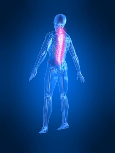
About The Spine
 The spine/vertebral column is a segmented flexible pillar. It makes up about two-fifths of the total height of the body. It is made up of a series of bones (vertebrae) which are separated by discs. The total number of vertebrae during early development is 33, then several vertebrae fuse, making 24 separate bones: 7 in the neck (cervical/neck vertebrae), designated C1 – C7, 12 in the chest/rib (thoracic vertebrae), designated T1 – 12, and 5 in the back (lumbar vertebrae), designated L1 – 5. They are all numbered from the top down so, the C1 vertebrae supports the skull – (If you like history – C1 is also called the “atlas” as the early anatomists thought it was like Atlas holding up the World - it is this joint that does most of head nodding up and down. They called C2 the “axis” as the joint between it and C1 allows most of the rotation movement of the head ie the shaking of your head from side to side – the owl movement).
Below the lumbar spine, lies the sacrum (the ‘sacred’ bone because it is rather cross shaped) which consists of five fused vertebral segments (S1 – 5) and a coccyx (tail bone) consisting of four tiny fused segments.
Did you know?
- The function of the vertebral column is essential to everything you do. It encloses the spinal cord, supports the head, the ribs and thereby allows you to breath, the pelvis and thereby the legs, the shoulder girdle and thereby the arms as well as all the spinal muscles which allow you to bend and twist your body and with your Atlas and Axis even communicate yes and no!
- The length of the column is about 71 cm in the average male and about 61 cm in an average female
- The spine has 4 normal gentle curves – the cervical and lumbar curves are convex forwards (bulge out), whereas the thoracic and sacral curves are concave forwards (cup in). The convex ones tend to make us lean backwards like a Lord and are called “lordosis” whilst the concave are called “kyphosis”. These normal lordotic and kyphotic curves are crucial to a healthy spine. The curves increase its strength, absorb shocks during movement and protect the vertebrae from fracture and the discs from wear and tear.
- We can lose these curves and develop a so called “flat back” or the curves may get exaggerated and get a hyper- kyphosis or lordosis or a sideways curve known as a scoliosis which lead to dysfunction in the spine.
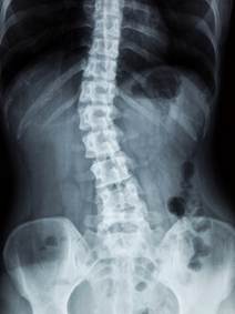
An X-ray of the spine showing a Scoliosis in the Lumbar Spine
The sacrum and coccyx are composed usually of fused bones but the rest of the spine is joined by individual articulations between the vertebrae, separated by discs (except between C1 and C2 vertebrae). These intervertebral discs are soft(ish) cushions which act as both shock absorbers and hinges. Each disc has an outer fibrous ring consisting of fibrocartilage called the annulus fibrosus (annulus = ring-like) and in inner soft, pulpy, highly elastic substance called the nucleus pulposus (pulposus = pulp-like). The discs form strong joints, permit various movements of the vertebral column and absorb vertical shock. Under compression, they flatten, broaden and bulge from their intervertebral spaces.
The physiology and mechanics of a disc is a superb feat of natural engineering. It has no natural blood supply and yet survives for an entire lifetime taking huge strains each time we bend, run or jump.
As the intervertebral discs play a large role in the spine, when they degenerate, they can cause huge problems such as disc prolapses/bulges.
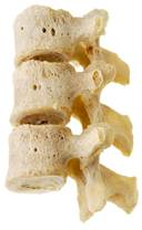
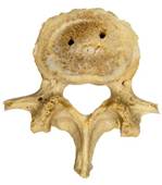
The vertebrae vary somewhat in size, shape and detail throughout the spine though the basic shape is as below.
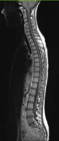
An MRI scan of the Whole Spine
Each vertebra forms a functional unit within the spine – “a motion segment”.
A Motion Segment consists of:
- The vertebral bodies/vertebrae
- An intervertebral disc
- A pair of facet joints (facets = little faces). About the size of little finger joints they are formed by the junction of bony processes, one each on the right and left, that interlock with those of the vertebrae above or below.
- In the cervical spine there are also small joints at the side of the discs called uncovertebral joints.
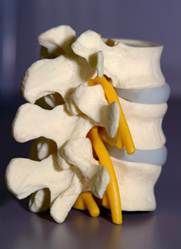
Human Facet Joints connecting the Lumbar Vertebrae
All the movements of the spine go through the discs and facet joints of these motion segments. If they become the subject of wear and tear they develop arthritis or spondylosis. If they become loose, as a result of this arthritis, one vertebra may slip on another – this slip is called a spondylolisthesis.
Pedicles project posteriorly from the body to unite with the laminae (meaning thin layers). A bony arch is formed, made up of the lamina and pedicles on each side and this is crowned by the spinous processes (the knobbly bit you can feel in the middle of your back where the fins stuck out on the back of dinosaurs.) This arch, together with the body of the vertebra, surrounds and protects the spinal cord.
A human lumbar vertebra with two transverse processes and a posterior spinous process
In addition, where the laminae and pedicle join, a transverse process extends. The spinous and transverse processes serve as points of attachment for muscles.
The spinal canal lies between the vertebral arch and body and contains the spinal cord, which is contained in a sack called the dura. This sack contains the cerebrospinal fluid (CSF). The spinal cord floats in the CSF and this protects it from the movements of the bones and discs of the spine. The canal also has a little fatty tissue and some blood vessels.
- A “lumbar puncture” is performed by passing a needle between two lamina, through the dura and into the CSF which is withdrawn for testing.
- A myelogram is performed by putting dye into the CSF and taking X-rays or scan pictures of the silhouettes made by the spinal cord or discs
- An epidural is performed by putting steroid or anaesthetic in the space between the dura and the bone of the lamina to relieve pain
- A “spinal anaesthetic” involves putting anaesthetic into the CSF to relieve pain.
The pedicles have little notches and when stacked up on one another, they form openings between the vertebrae on both sides of the vertebral column – each opening (intervertebral foramen), permits the passage of a single spinal nerve root.
A model showing the emerging spinal nerve roots, leaving the spinal canal at the intervertebral foramina
Cervical, thoracic and lumbar spinal nerve roots pass through the intervertebral foramina and can get trapped by bulging discs to cause arm, chest or leg pain/weakness/pins and needles.
The brain is connected to the spinal cord through the foramen magnum in the base of the skull - (Foramen magnum means “big hole”. The base of the skull also has lots of smaller holes through which nerves to the nose and tongue pass to serve smell and taste, and other holes for the blood vessels that take blood to and from the brain)
The spinal cord contains about 100 million neurons and is essentially the brain’s motorway, linking it to all the trunk roads or spinal nerve roots that come off the spinal cord at each motion segment. These nerve roots exit the spinal canal via the intervertebral foramen to go down the arms and legs. These trunk roads/roots divide into smaller and smaller nerves as they pass through the body to reach even the remotest outer reaches of your body. This network of roads is called the “peripheral nerves”.
Taken together the brain, spinal cord and peripheral nerves make up the “nervous system”. The spinal cord and brain alone are sometimes referred to as the “central nervous system”.
The spinal cord is soft, about the size of a finger and greyish in colour. Thirty-one pairs of spinal nerve roots emerge from the spinal cord, each serving a specific region of the body. The spinal cord ends at the top of the lumbar spine which lies approximately level with the naval.
In the lumbar spinal canal, there is no spinal cord but lots of nerve roots which supply the functions of the legs (power and feeling), the bowel and bladder.
The spinal cord contains all of the nerves controlling feeling and movement of every part of the body along with all of the conscious and unconscious actions of the internal organs, chest and abdomen.
Did you know: Though made up of billions of nerves, the spinal cord is entirely numb? However, the spinal nerve roots are not; when a disc touches a nerve root – it can hurt like fury, as felt by anyone who has had a lumbar disc prolapse.